Tendon Anatomy
Tendons act as links between muscle and bone, enabling force to be transmitted and movement to occur at the joints. Each muscle contains two tendons, one proximal and one distal. The point at which the tendon joins the muscle is known as the musculotendinous junction, while the area where the tendon joins the bone is referred to as the osteotendinous junction (Jonsson 2009). On observation healthy tendons are a white colour with a fibro elastic texture. Tendons are covered by an epitenon which is, in turn, surrounded by a paratenon. The epitenon is a fine, loose connective-tissue sheath through which blood vessels, lymphatics and nerves are all contained (Jonsson 2009). The paratenon is described as being a loose areolar connective tissue covering mainly comprised of type 1 and 3 collagen fibrils, some elastic fibrils and an inner lining of synovial cells (Sharma and Maffuli 2005).
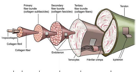
Fig 1.1: Diagrammatic representation of Tendon composition (Bjur 2010).
Tendons comprise of a number of basic elements, which include: collagen bundles, cells and ground substance. Tensile strength of tendons is attributed to the collagen formation within. This collagen formation may be primarily longitudinal, horizontal, or transverse in nature depending on the direction of load which the tendon commonly absorbs. The ground substance provides structural support to the collagen fibres (Sharma and Maffuli 2005). Normal tendons consist of dense, parallel and slightly wavy collagen bundles which display a reflective appearance under polarised light (Sharma and Maffuli 2005).
Tendons are composed, in the majority, by cells known as tenocytes. These cells are fibroblastic in nature; meaning they synthesise the formation of collagen and extracellular matrix (ECM) (Rio et al 2013). The collagen and ground substance referenced earlier is all encapsulated within the ECM and tenocytes lie end to end in channels between the collagen. Communication between these cells, in relation to environmental conditions, is regulated by gap junctions (Rio et al 2013). Calcium concentration, pH and mechanical load all influence the opening/closing patterns of gap junctions and, in turn, the activity of tenocytes.
Blood supply to the tendon is provided by a network of small arterioles which are orientated parallel to the collagen fibres within the endotenon (Cook et al 1998). Despite this blood supply tendons have been reported as displaying approximately 8 times lower O2 consumption when compared to skeletal muscle (Jonsson 2009). The benefit of this is that the tendon can remain under load and tension for longer without risk of ischaemia and injury. However this low O2 consumption also may play a role in impaired and slower healing after injury (Maffuli et al 2009).